Macronodular liver cirrhosis with ascites
Written and reviewed by RadLearn editorial team · Updated 20 Oct 2026
Figures (10)
Patient history
The patient presented with weakness, loss of appetite, dizziness and dyspnoea. Her skin had become dry and brown. Blood tests showed anaemia with raised ALT, AST, urea and creatinine. Palpation revealed a firm, non-tender abdomen. On examination the abdomen appeared as though she were eight months pregnant.
Imaging findings — key points
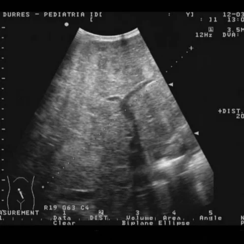
- Ultrasound of the liver shows a coarse, heterogeneous parenchyma with a nodular surface and multiple large regenerative nodules, together with a large volume of ascites.
- The left lobe is disproportionately enlarged, and the spleen is enlarged with a heterogeneous echotexture, while both kidneys show increased cortical echogenicity.
- Axial CT demonstrates a nodular hepatic contour with macronodules of varying size that are isodense to the surrounding parenchyma in all phases, without arterial hyperenhancement or washout.
- There is a large amount of ascites with peritoneal enhancement and scalloping of the liver surface, and the spleen is enlarged with varices in the splenic hilum.
- No enhancing focal lesion, portal vein thrombosis or biliary dilatation is identified.
Discussion
Macronodular cirrhosis is the end stage of chronic liver injury in which broad fibrous septa surround large regenerative nodules, and it may be caused by viral hepatitis, alcohol, autoimmune disease or metabolic disorders. Imaging shows a nodular liver contour, coarse parenchyma, splenomegaly, ascites and portosystemic collaterals, and the main diagnostic challenge is separating benign regenerative nodules from hepatocellular carcinoma, which arises in this background at an annual risk of several per cent. Regenerative nodules are typically isodense or isointense to liver on all phases and do not restrict diffusion, whereas HCC shows arterial enhancement with washout. Surveillance with ultrasound and, when necessary, contrast-enhanced CT or MRI is essential for early detection of malignant transformation.
Differential diagnosis
| Alternative diagnosis | Key features that distinguish it from this case |
|---|---|
| Hepatocellular carcinoma | Hepatocellular carcinoma shows arterial hyperenhancement with portal venous or delayed washout and may invade the portal vein, whereas regenerative macronodules remain isodense in all phases. |
| Fibrolamellar carcinoma | Fibrolamellar carcinoma occurs in young patients without cirrhosis, is a large heterogeneous mass with a central scar and calcification, and does not produce a diffusely nodular liver. |
| Multifocal hepatocellular carcinoma | Multifocal HCC produces discrete enhancing nodules with washout and tumour thrombus, unlike the diffuse, non-enhancing regenerative nodules of macronodular cirrhosis. |
| Hepatic metastasis | Metastases are usually multiple, hypodense and rim-enhancing with a known primary, and they do not follow the diffuse nodular contour of cirrhosis. |
The confirmed diagnosis itself is not included as an alternative — it is stated separately below.