Le Fort III fracture (craniofacial disjunction)
Written and reviewed by RadLearn editorial team · Updated 20 Oct 2026
Figures (7)
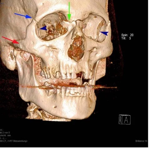
Patient history
A 50-year-old man was brought to the emergency department by ambulance after a fall onto his face, with extreme swelling and multiple bruises and haematomas, raising the possibility of a midface fracture.
Imaging findings — key points
- 3D CT reconstruction of the skull shows a craniofacial disjunction, with fracture lines passing through the frontonasal suture, the medial orbital walls, the zygomaticofrontal sutures and the zygomatic arches, separating the entire facial skeleton from the cranial base.
- Axial unenhanced CT of the paranasal sinuses demonstrates fractures of the pterygoid plates and disruption of the pterygomaxillary junction, with air and haematoma in the pterygopalatine fossa and infratemporal fossa.
- Coronal reformats confirm a detached midface with depression of the malar eminences, haemosinus and extensive subcutaneous emphysema of the face and orbits.
- The 3D view from below confirms that the maxilla and zygomas move as a unit with the cranium, in contrast with the lower Le Fort types.
- There is no intracranial haemorrhage, pneumocephalus or displaced cranial vault fracture on the available images.
Discussion
Le Fort fractures are classified by the level at which the facial skeleton separates from the cranial base, and type III is the craniofacial disjunction in which the fracture line runs through the nasofrontal suture, the medial orbital walls and the zygomatic arches. Clinically the patient has a mobile, depressed midface, periorbital haematoma and often a cerebrospinal fluid leak, and airway protection takes priority over imaging. CT with thin slices and 3D reconstruction is the definitive investigation and allows the radiologist to map every component of the injury, including pterygoid plate involvement, orbital floor disruption and associated intracranial trauma. Accurate classification guides surgical fixation and predicts which patients need neurosurgical and ophthalmological review.
Differential diagnosis
| Alternative diagnosis | Key features that distinguish it from this case |
|---|---|
| Le Fort II fracture | A Le Fort II fracture is pyramidal and passes through the nasofrontal suture, medial orbital walls and infraorbital rims but spares the zygomaticofrontal sutures and zygomatic arches, so the zygomas remain attached to the cranium. |
| Le Fort I fracture | A Le Fort I fracture is a horizontal fracture through the maxilla above the tooth apices with pterygoid plate involvement, leaving the orbit and zygomas intact. |
| Zygomaticomaxillary complex fracture | A zygomaticomaxillary complex fracture involves the zygomatic arch, lateral orbital wall and orbital floor on one side and produces malar depression without a mobile whole midface. |
The confirmed diagnosis itself is not included as an alternative — it is stated separately below.