Duplicated median nerve
Written and reviewed by RadLearn editorial team · Updated 20 Oct 2026
Figures (3)
Patient history
The authors describe a case of a duplicated median nerve associated with carpal tunnel syndrome.
Imaging findings — key points
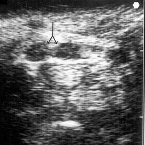
- Axial ultrasound of the wrist shows two separate nerve fascicles within the carpal tunnel, each with a fascicular echotexture, lying side by side and separated by a thin fascial septum.
- The median nerve is enlarged and hypoechoic at the level of the carpal tunnel, consistent with compression neuropathy, and the flexor retinaculum is bowed.
- Axial T1-weighted MRI demonstrates a bifid median nerve with two nerve bundles within the carpal tunnel, and no space-occupying lesion is present.
- There is no persistent median artery, ganglion, lipoma or tenosynovial proliferation causing the compression.
- The thenar muscles show no fatty atrophy on the available images.
Discussion
Duplication of the median nerve is a rare anatomical variant in which the nerve splits within the forearm or carpal tunnel, and it is clinically important because each branch may be compressed separately and because it increases the risk of iatrogenic injury during carpal tunnel release. Ultrasound and MRI show two discrete nerve bundles within the tunnel, sometimes separated by a persistent median artery, and the nerve may be enlarged and hypoechoic from compression. The differential includes a nerve sheath tumour and tenosynovitis, both of which produce a mass rather than a duplicated nerve. Preoperative recognition allows the surgeon to decompress both branches and avoid incomplete release.
Differential diagnosis
| Alternative diagnosis | Key features that distinguish it from this case |
|---|---|
| Bifid median nerve with persistent median artery | A persistent median artery lies between the two nerve bundles and shows a Doppler signal, whereas in this case the duplication is present without a central artery. |
| Median nerve schwannoma | A schwannoma is a focal enhancing mass that displaces the nerve and does not show two discrete fascicular bundles with a normal nerve texture. |
| Flexor tenosynovitis | Tenosynovitis produces fluid and synovial thickening around the flexor tendons, compressing a single median nerve rather than creating a duplicated nerve. |
| Normal variant median nerve | A normal median nerve is a single bundle without a longitudinal septum or two separate epineurial sheaths, as seen here. |
The confirmed diagnosis itself is not included as an alternative — it is stated separately below.