Inflammatory breast cancer
Written and reviewed by RadLearn editorial team · Updated 20 Oct 2026
Figures (10)
Patient history
A 35-year-old woman complained of right breast pain for two months. She received antibiotic treatment without improvement. On physical examination, the right breast was enlarged, erythematous, and warm. Nipple retraction was present. A palpable breast mass and two fixed ipsilateral axillary lymph nodes were also detected.
Imaging findings — key points
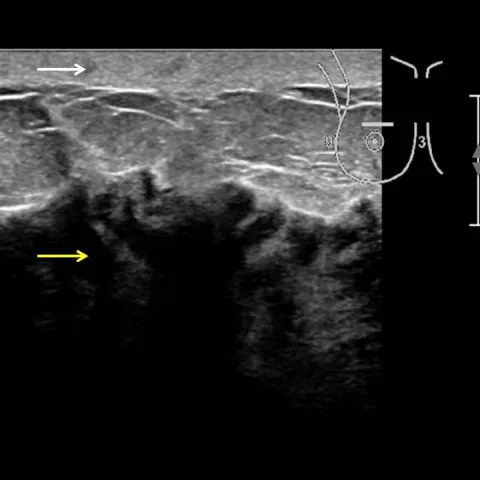
- Ultrasound shows a diffuse hypoechoic region throughout the enlarged right breast, with skin thickening and nipple retraction.
- Ultrasound identifies four abnormal right axillary lymph nodes, while the contralateral breast and axilla are normal.
- Dynamic contrast-enhanced breast MRI shows irregular, heterogeneous, asymmetric non-mass enhancement involving a large portion of the right breast, with a type 2 (plateau) kinetic curve.
- MRI also shows thickening of the right pectoralis muscle, consistent with oedema of the anterior chest-wall space.
- Mammography could not be obtained because of severe breast pain, so no mammographic assessment was possible.
- Staging CT demonstrates multiple bone metastases, corresponding to a final stage of T4dN2M1.
Discussion
Inflammatory breast cancer is a rare and highly aggressive form of locally advanced breast cancer characterised by diffuse skin erythema and oedema (peau d'orange), and its diagnosis rests on a rapid onset of typical symptoms, a history shorter than six months, erythema involving at least one third of the breast, and histopathological confirmation of invasive carcinoma. Biopsy in this case showed an invasive ductal carcinoma with an aggressive phenotype (HER2-negative, ER 32%, PR 0%, Ki67 44%), and skin punch biopsy may demonstrate dermal lymphatic tumour emboli. Because approximately 30% of patients have distant disease at presentation, staging with CT and bone scanning is mandatory; management is neoadjuvant chemotherapy followed by mastectomy and post-mastectomy radiotherapy, and breast-conserving surgery with sentinel node biopsy is inappropriate, with prognosis remaining poor.
Differential diagnosis
| Alternative diagnosis | Key features that distinguish it from this case |
|---|---|
| Acute infectious mastitis or breast abscess | Presents with fever, leukocytosis and focal tenderness with a drainable collection, and improves within one to two weeks of antibiotics, whereas inflammatory breast cancer shows no response. |
| Locally advanced non-inflammatory breast carcinoma | Presents as a dominant mass with overlying skin changes but lacks the diffuse erythema and dermal oedema occupying more than one third of the breast that defines inflammatory disease. |
| Granulomatous mastitis | Shows lobulocentric granulomas on biopsy without malignant cells or dermal lymphatic tumour emboli, and follows a chronic rather than rapidly progressive course. |
| Radiation dermatitis or radiation recall | Is confined to a previously irradiated field, resolves with supportive care, and produces no diffuse breast enlargement or dermal lymphatic involvement. |
The confirmed diagnosis itself is not included as an alternative — it is stated separately below.