Infective sacroiliitis complicated by a pelvic muscle abscess
Written and reviewed by RadLearn editorial team · Updated 20 Oct 2026
Figures (7)
Patient history
A 40-year-old man presented with left flank pain and tenderness radiating to the left gluteal region, with night sweats and rigors over the previous 48 hours. He reported occasional intravenous cocaine use. Laboratory tests: CRP 58.5 mg/l and ESR 92 mm.
Imaging findings — key points
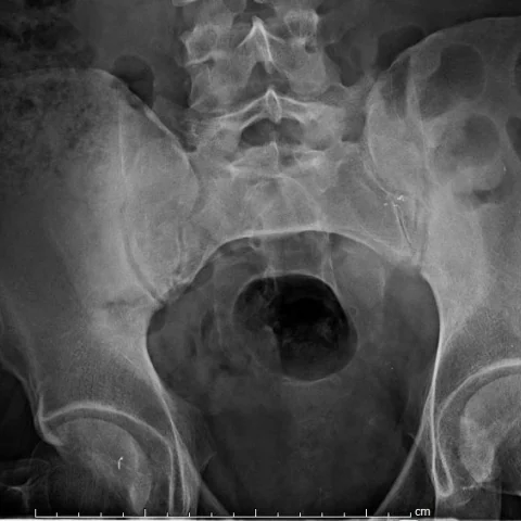
- The AP radiograph of the pelvis shows widening of the left sacroiliac joint with irregularity of the adjacent articular margins and mild surrounding sclerosis.
- CT in the bone window demonstrates erosions on both the iliac and sacral sides of the left sacroiliac joint, with subchondral sclerosis and joint space widening.
- Contrast-enhanced pelvic CT shows a rim-enhancing fluid collection within the left iliacus and piriformis muscles, with surrounding fat stranding and muscle swelling.
- There is no gas within the joint or soft tissue, and the right sacroiliac joint appears normal.
- The collection extends along the pelvic sidewall and exerts mass effect on the adjacent vessels without thrombosis.
Discussion
Infective sacroiliitis is an uncommon but serious infection that usually results from haematogenous spread, often in patients with intravenous drug use, immunosuppression or a distant source, and Staphylococcus aureus is the most frequent organism. The infection begins in the synovium and spreads to the subchondral bone, producing erosions, joint widening and eventually a pelvic muscle abscess. CT and MRI are the key investigations, with contrast-enhanced CT or MRI showing joint effusion, bone marrow oedema, erosions and rim-enhancing collections in the iliacus or piriformis muscle. Prompt antibiotic therapy and drainage of the abscess are essential, and delay can lead to septic arthritis, bone destruction and sepsis.
Differential diagnosis
| Alternative diagnosis | Key features that distinguish it from this case |
|---|---|
| Degenerative sacroiliitis | Degenerative disease produces osteophytes, subchondral sclerosis and vacuum phenomenon with a preserved joint space in older patients, without erosions, fever or a soft-tissue abscess. |
| Inflammatory sacroiliitis | Inflammatory sacroiliitis is usually bilateral and symmetric in a young patient with inflammatory back pain and HLA-B27 association, without a fluid collection or systemic infection. |
| Infective sacroiliitis without abscess | Early infection may show only joint effusion and erosions, but the presence of a rim-enhancing pelvic muscle collection indicates complicated disease beyond the joint. |
| Sacral insufficiency fracture | An insufficiency fracture shows a sclerotic band or fracture line parallel to the sacroiliac joint without erosions, joint widening or an abscess. |
The confirmed diagnosis itself is not included as an alternative — it is stated separately below.