Hydroxyapatite deposition disease (HADD), intra-articular
Written and reviewed by RadLearn editorial team · Updated 20 Oct 2026
Figures (4)
Patient history
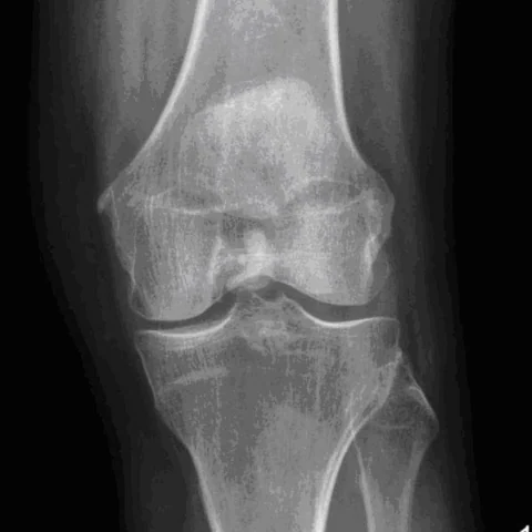
A 72-year-old woman presented with a six-month history of left knee pain that was not relieved by anti-inflammatory medication. Physical examination found a moderate joint effusion with medial joint line tenderness. At follow-up three weeks later she was completely unable to flex or extend the left knee.
Imaging findings — key points
- The digital radiograph of the left knee shows amorphous, cloud-like calcification in the periarticular soft tissues and within the joint recesses, without bone erosion or fracture.
- Sagittal, axial and coronal fat-saturated T2-weighted MRI show multiple low-signal calcific deposits within the synovium and joint fluid, surrounded by a moderate joint effusion and synovial thickening.
- The calcific masses are intra-articular and extend into the suprapatellar and posterior recesses, producing mechanical restriction of movement.
- There is no soft-tissue mass with contrast enhancement and no marrow abnormality.
- A follow-up radiograph shows progression of the calcification with worsening joint stiffness and inability to flex or extend the knee.
Discussion
Hydroxyapatite deposition disease results from the deposition of basic calcium phosphate crystals in periarticular tissues, most often the rotator cuff, but intra-articular involvement of the knee can occur and produce acute or chronic pain, effusion and mechanical restriction. Imaging shows amorphous, cloud-like calcification in the synovium or joint capsule, with low signal on all MRI sequences and an associated effusion, and the deposits may be resorbed spontaneously. The main differentials are synovial sarcoma, CPPD and gout, each of which has a different pattern of calcification and bone change. Treatment is supportive with analgesia, aspiration and physiotherapy, and surgical removal is reserved for refractory mechanical symptoms.
Differential diagnosis
| Alternative diagnosis | Key features that distinguish it from this case |
|---|---|
| Synovial sarcoma | Synovial sarcoma is a soft-tissue mass that enhances and may calcify, but it arises adjacent to the joint rather than forming multiple intra-articular calcific bodies within synovium. |
| Calcium pyrophosphate deposition disease | CPPD produces punctate or linear chondrocalcinosis in the menisci and hyaline cartilage, not large amorphous periarticular calcific masses. |
| Gout | Gout shows punched-out juxta-articular erosions with overhanging edges and tophi of intermediate signal, and the calcification is not typically cloud-like and intra-articular. |
| Synovial chondromatosis | Synovial chondromatosis produces multiple well-defined osteochondral bodies with ring-and-arc calcification and marrow signal, rather than amorphous hydroxyapatite deposits. |
The confirmed diagnosis itself is not included as an alternative — it is stated separately below.