Gastric pneumatosis due to gastric outlet obstruction
Written and reviewed by RadLearn editorial team · Updated 20 Oct 2026
Figures (7)
Patient history
A 42-year-old man was admitted with postprandial abdominal pain, bloating, nausea and vomiting. He had a history of epigastric pain for about five months. He had undergone gastroscopy a few days earlier. Clinical examination showed distension and mild tenderness without rigidity. The remainder of the examination was unremarkable.
Imaging findings — key points
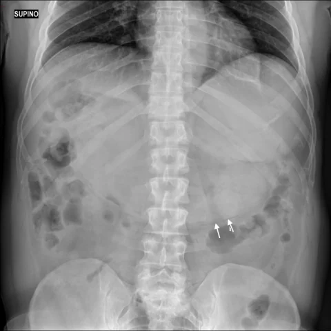
- The abdominal radiograph shows a markedly distended stomach with a large air-fluid level and a mottled gas pattern, and there is linear gas within the gastric wall.
- Multidetector CT demonstrates severe gastric distension with retained food and fluid, and circumferential intramural gas in the stomach wall.
- The stomach wall is otherwise thin without marked thickening or enhancement abnormality, and there is no extraluminal free gas or portal venous gas.
- There is a transition point at the gastric outlet with narrowing of the pylorus or duodenum, consistent with outlet obstruction.
- No pneumoperitoneum, ascites or mesenteric stranding is identified.
Discussion
Gastric pneumatosis is the presence of gas within the gastric wall, and it has a benign and a life-threatening cause; when it occurs distal to a gastric outlet obstruction it is usually reversible once the obstruction is relieved. CT is essential to distinguish intramural gas from free intraperitoneal gas, to identify the transition point and to assess for portal venous gas or wall thickening that would suggest emphysematous gastritis. The absence of sepsis, wall necrosis and portal venous gas supports a benign mechanism related to raised intraluminal pressure. Management is directed at the obstruction, with nasogastric decompression and endoscopic or surgical relief as required.
Differential diagnosis
| Alternative diagnosis | Key features that distinguish it from this case |
|---|---|
| Pneumoperitoneum | Pneumoperitoneum shows free gas outside the bowel lumen, often under the diaphragm, rather than gas confined within the gastric wall. |
| Gastric emphysema | Gastric emphysema is intramural gas without obstruction or severe illness that resolves with conservative treatment, whereas this case shows a clear outlet obstruction causing the pneumatosis. |
| Emphysematous gastritis | Emphysematous gastritis is a life-threatening infection with marked wall thickening, fluid, sepsis and gas dissecting into the portal venous system, unlike the thin-walled distended stomach here. |
| Gastric volvulus | Gastric volvulus produces a distended, twisted stomach with a bird-beak transition and may cause ischaemia, but the classic double-bubble or organo-axial configuration is absent here. |
The confirmed diagnosis itself is not included as an alternative — it is stated separately below.