Ectopic nasal tooth
Written and reviewed by RadLearn editorial team · Updated 20 Oct 2026
Figures (2)
Patient history
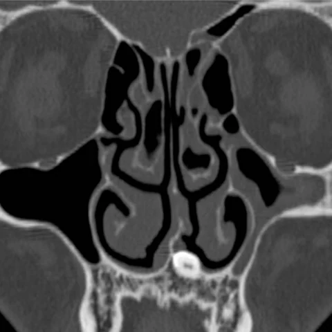
A 33-year-old man had a history of recurrent sinusitis and nasal discharge. Clinical examination found a hard mass in the floor of the left nasal cavity.
Imaging findings — key points
- Coronal high-resolution CT of the paranasal sinuses shows a well-defined, markedly radiodense structure lying in the floor of the left nasal cavity, with an outer layer of enamel density and a central low-density pulp cavity.
- The structure has the morphology of a tooth, with a crown and root, and is surrounded by a thin bony rim continuous with the maxillary alveolar process.
- There is mucosal thickening of the adjacent left maxillary sinus and nasal cavity, consistent with recurrent sinusitis, but no bone destruction or aggressive periosteal reaction.
- The maxillary dental arch is otherwise intact, and no other ectopic or supernumerary teeth are seen.
- No laminated calcification or foreign-body density is present to suggest a rhinolith.
Discussion
An ectopic nasal tooth is a rare developmental anomaly in which a tooth erupts into the nasal cavity instead of the oral cavity, most often through the floor of the nose or the anterior maxilla. It may be asymptomatic or present with unilateral nasal obstruction, recurrent sinusitis, epistaxis or facial pain, and the diagnosis is usually made on CT when a tooth-like structure with enamel and a pulp cavity is identified. The main differential is a rhinolith, which is a laminated calcified concretion without dental architecture, and a foreign body. Treatment is endoscopic removal, which is curative and avoids damage to adjacent structures.
Differential diagnosis
| Alternative diagnosis | Key features that distinguish it from this case |
|---|---|
| Rhinolith | A rhinolith is a laminated, irregular calcified mass formed around a nidus, lacks enamel and a pulp cavity, and is usually centred in the nasal airway rather than attached to the alveolar bone. |
| Foreign body | A foreign body has a characteristic density and shape of the ingested material, a clear history of insertion and no tooth-like crown or root architecture. |
| Odontoma | An odontoma is a hamartomatous mass of dental tissues within the jaws, often with multiple denticles, and it does not lie free in the nasal floor. |
| Calcified nasal polyp | A calcified polyp arises from the nasal mucosa, shows soft-tissue density with scattered calcification and is not surrounded by a bony follicle. |
The confirmed diagnosis itself is not included as an alternative — it is stated separately below.