Cutaneous Scalp Metastasis Secondary to Lung Cancer (NSCLC)
Written and reviewed by RadLearn editorial team · Updated 22 Oct 2026
Figures (11)
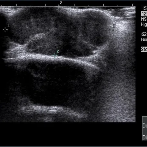
Patient history
A 67-year-old man, diagnosed 5 months earlier with lung cancer (non-small cell, T4N0M0) and treated with palliative radiotherapy, was referred by his primary care physician for investigation of a 30 mm skin/subcutaneous mass in the occipital region of the scalp with superficial erosion. He had a history of prostate cancer.
Imaging findings — key points
- Cutaneous metastases from lung cancer are uncommon but may present as a scalp mass.
- A 30 mm skin/subcutaneous occipital mass with superficial erosion was demonstrated on ultrasound.
- Colour Doppler ultrasound was used to assess the lesion.
- Non-contrast CT was performed to evaluate the occipital lesion.
- Biopsy confirmed the diagnosis of metastasis secondary to lung cancer (NSCLC).
- A history of lung cancer should raise suspicion for cutaneous metastasis when a new scalp lesion appears.
Discussion
This case illustrates an unusual presentation of cutaneous metastasis from lung cancer (NSCLC) to the scalp. Cutaneous metastases are rare but should be considered when a new skin/subcutaneous mass appears in a patient with a known malignancy, particularly when superficial erosion is present. Ultrasound with colour Doppler and non-contrast CT help characterise the lesion and define its extent, but tissue sampling is required for confirmation. The main pitfalls are misdiagnosing the lesion as a benign cyst or lipoma and failing to obtain a biopsy in a patient with a history of lung cancer. Management implication: recognition of cutaneous metastasis alters staging and may prompt adjustment of palliative therapy.
Differential diagnosis
| Alternative diagnosis | Key features that distinguish it from this case |
|---|---|
| Epidermal inclusion cyst | Usually well-defined and cystic without superficial erosion; biopsy excludes it. |
| Sebaceous cyst | Typically cystic and slow-growing; erosion and history of lung cancer argue against it. |
| Lipoma | Fat-density on CT and avascular on Doppler; not expected to erode the skin. |
| Haemangioma | Shows vascular flow on colour Doppler and lacks the clinical context of lung cancer. |
The confirmed diagnosis itself is not included as an alternative — it is stated separately below.