Chronic eosinophilic pneumonia
Written and reviewed by RadLearn editorial team · Updated 20 Oct 2026
Figures (7)
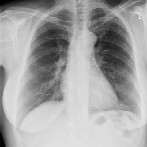
Patient history
A 54-year-old woman attended the outpatient asthma clinic with a several-month history of cough, fatigue, shortness of breath on minimal exertion and wheeze. Blood tests showed a raised erythrocyte sedimentation rate and eosinophilia of 2.0 x 10^9/L. There was no fever.
Imaging findings — key points
- The chest radiograph at presentation shows bilateral, predominantly peripheral airspace opacification in the mid and upper zones, producing a photographic negative of pulmonary oedema.
- CT and HRCT demonstrate patchy peripheral and subpleural consolidation with ground-glass opacity, sparing the central perihilar regions.
- There is no significant pleural effusion, lymphadenopathy, cavitation or honeycombing, and the airways are patent.
- A radiograph taken after clinical deterioration shows progression of the peripheral consolidation, and the post-treatment HRCT shows complete or near-complete resolution after corticosteroid therapy.
- The distribution and rapid resolution are characteristic of an eosinophilic airspace process rather than infection or fibrosis.
Discussion
Chronic eosinophilic pneumonia is a rare idiopathic eosinophilic lung disease that occurs most often in middle-aged women, frequently with a background of asthma, and presents with cough, dyspnoea, wheeze and peripheral blood eosinophilia. The characteristic radiographic pattern is bilateral peripheral consolidation that spares the perihilar regions and resembles the photographic negative of pulmonary oedema, and HRCT confirms subpleural consolidation and ground-glass opacity. The diagnosis is supported by bronchoalveolar lavage eosinophilia and by rapid clinical and radiological response to corticosteroids, which is also the treatment. Relapse after withdrawal is common, so a prolonged tapering course and follow-up imaging are required.
Differential diagnosis
| Alternative diagnosis | Key features that distinguish it from this case |
|---|---|
| Drug-related pulmonary infiltrates | Drug-induced eosinophilic pneumonia follows a newly started medication and usually resolves after withdrawal, whereas this case has no drug trigger and recurs without steroids. |
| Allergic bronchopulmonary aspergillosis | ABPA shows central bronchiectasis, mucoid impaction and high IgE with Aspergillus sensitisation, rather than peripheral consolidation without bronchial dilatation. |
| Parasitic infection | Parasitic pulmonary eosinophilia follows travel to an endemic area and may show migratory infiltrates, and it is associated with specific serology. |
| Cryptogenic organising pneumonia | Organising pneumonia also responds to steroids but typically shows peribronchovascular or subpleural consolidation with reverse halo signs, and peripheral eosinophilia is absent. |
The confirmed diagnosis itself is not included as an alternative — it is stated separately below.