Acute disseminated encephalomyelitis (ADEM)
Written and reviewed by RadLearn editorial team · Updated 20 Oct 2026
Figures (18)
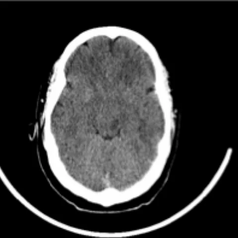
Patient history
An 18-year-old man was admitted to the emergency department with a five-day neurological illness consisting of motor impairment, clumsiness of the right hand and dysarthria. In the preceding days he had a productive cough without fever. Lumbar puncture, blood tests and cerebrospinal fluid were normal. Antibody testing was also negative.
Imaging findings — key points
- Unenhanced CT of the brain is essentially normal or shows only ill-defined, asymmetric hypodensity in the deep white matter, without haemorrhage or mass effect.
- Axial FLAIR images demonstrate multiple bilateral but asymmetric hyperintense lesions in the subcortical and deep white matter, with poorly defined margins and a tendency to become confluent.
- Axial T2-weighted images confirm the lesions, which involve the centrum semiovale and may extend to the brainstem, cerebellum, thalami and basal ganglia, with mild surrounding oedema but no significant mass effect.
- Contrast-enhanced T1-weighted images show variable enhancement, frequently ring-like or open-ring enhancement in larger lesions, and no meningeal enhancement.
- Follow-up MRI five months later shows marked regression or complete resolution of the lesions with residual gliosis, supporting a monophasic demyelinating process rather than multiple sclerosis.
Discussion
Acute disseminated encephalomyelitis is an immune-mediated demyelinating disease that typically follows a viral infection or vaccination by one to three weeks and is more common in children and young adults. Lesions are multifocal, asymmetric and poorly demarcated, affecting the white matter and frequently the deep grey matter, brainstem and cerebellum; the perivenular demyelination explains the small size of individual lesions and their tendency to coalesce. The key clinical question is separation from a first attack of multiple sclerosis, and the combination of a preceding infection, encephalopathy, large poorly marginated lesions and complete or near-complete resolution on follow-up imaging favours ADEM. Treatment is high-dose corticosteroids, with plasma exchange or intravenous immunoglobulin in refractory cases.
Differential diagnosis
| Alternative diagnosis | Key features that distinguish it from this case |
|---|---|
| Multiple sclerosis | Multiple sclerosis produces smaller periventricular ovoid lesions perpendicular to the ventricles, often with chronic black holes, and new lesions appear over time, whereas ADEM is monophasic with large, poorly marginated lesions that resolve. |
| Epstein-Barr virus encephalitis | EBV encephalitis usually follows a febrile illness with lymphadenopathy and shows more symmetric involvement of the basal ganglia and thalami, often with meningeal enhancement. |
| Autoimmune paraneoplastic encephalitis | Autoimmune encephalitis tends to involve the limbic system with bilateral medial temporal hyperintensity and is associated with positive neuronal antibodies and a subacute neuropsychiatric course. |
| Bihemispheric diffuse glioma | A diffuse glioma produces progressive, expansile T2 hyperintensity with mass effect, altered perfusion and no spontaneous regression on follow-up, unlike the resolving lesions of ADEM. |
The confirmed diagnosis itself is not included as an alternative — it is stated separately below.